
Advanced Anatomy and Physiology: Fluid Balance
Prehospital Academy Lesson 7.8
This lesson reinforces and expands upon the lecture on fluid balance physiology. For EMTs, fluid balance is foundational to understanding shock, dehydration, edema, electrolyte disturbances, and the safe selection of intravenous fluids. Fluid balance underpins perfusion, blood pressure, cellular function, and neurologic status.
Why Fluid Balance Matters
Humans are largely composed of water, ranging from approximately 75 percent of body mass in infants to about 50 to 60 percent in adults, and even lower in older adults. Maintaining appropriate fluid balance is essential to homeostasis, the stable internal environment that allows cells, tissues, and organs to function effectively.
Adequate hydration and precise regulation of fluid levels support temperature regulation, nutrient transport, waste removal, and blood pressure maintenance. Disruption of fluid balance can lead to dehydration, electrolyte abnormalities, impaired perfusion, and life-threatening complications.
How the Body Maintains Fluid Balance
The body regulates fluid balance through several mechanisms, including thirst, hormonal control, and renal filtration. Thirst drives voluntary fluid intake, while the kidneys continuously adjust water and electrolyte excretion to match physiologic needs.
When these mechanisms fail or are overwhelmed by illness, injury, or environmental exposure, fluid imbalances develop. For EMTs, recognizing early signs of imbalance allows timely intervention and prevents deterioration.
Major Fluid Compartments of the Body
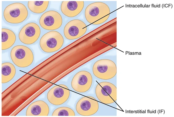
Fluid compartments are regions of the body separated by physical or functional barriers. The two primary compartments are intracellular fluid and extracellular fluid.
Intracellular fluid is contained within cells and makes up roughly 60 percent of total body water. It forms the cytoplasm and supports all intracellular metabolic processes.
Extracellular fluid surrounds cells and includes plasma and interstitial fluid. Plasma circulates within blood vessels and transports cells, proteins, electrolytes, nutrients, gases, and waste products. Interstitial fluid bathes cells and facilitates exchange between capillaries and tissues.
Specialized Extracellular Fluids
In addition to plasma and interstitial fluid, the body contains specialized extracellular fluids. These include cerebrospinal fluid, which protects the brain and spinal cord; lymph, which supports immune and fluid balance; pleural fluid in the thoracic cavities; and peritoneal fluid in the abdominal cavity.
Although these fluids are smaller in volume, disturbances can have serious clinical consequences, such as increased intracranial pressure or impaired respiration.
Cell Membranes and Selective Permeability
Cell membranes are selectively permeable. They allow lipid-soluble substances to cross freely while restricting many water-soluble substances. To sustain life, cells rely on transport mechanisms that regulate what enters and exits.
Movement across membranes occurs along concentration gradients or against them using energy. These processes maintain appropriate fluid and solute balance between compartments.
Diffusion and Active Transport
Diffusion is the passive movement of molecules from areas of higher concentration to lower concentration. It does not require energy. Oxygen and carbon dioxide move by diffusion between alveoli and pulmonary capillaries.
Active transport moves substances against their concentration gradients and requires energy in the form of ATP. A key example is the sodium-potassium pump, which maintains electrical and chemical gradients essential for nerve conduction and muscle contraction.
Filtration and Hydrostatic Pressure
Filtration uses pressure gradients to move fluid and solutes across membranes. In capillaries, hydrostatic pressure pushes plasma and nutrients into surrounding tissues. This process supplies cells with essential substances.
Oncotic pressure, also called colloid osmotic pressure, pulls fluid back into capillaries. At the arteriolar end of capillaries, hydrostatic pressure exceeds oncotic pressure, promoting fluid exit. At the venous end, oncotic pressure exceeds hydrostatic pressure, drawing fluid back in.
Filtration in the Kidneys
Filtration is also essential in the kidneys, where hydrostatic pressure drives blood filtration in the nephrons. This process produces filtrate that will ultimately become urine after reabsorption and secretion.
Disruption of renal filtration can rapidly affect fluid balance, blood pressure, and electrolyte levels.
Osmosis: Movement of Water
Osmosis is the diffusion of water across a semi-permeable membrane from areas of higher water concentration to lower water concentration. Water moves to balance solute concentrations between compartments.
Osmosis drives fluid shifts between intracellular and extracellular compartments and plays a central role in cell volume regulation.
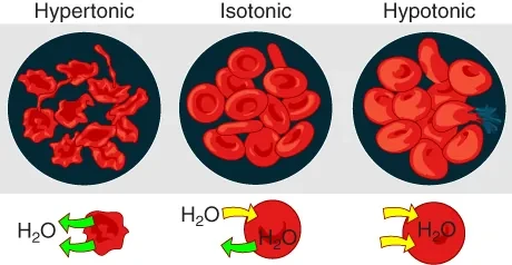
Tonicity: Isotonic, Hypotonic, and Hypertonic
Tonicity describes the relative concentration of solutes between two solutions. An isotonic solution has the same solute concentration as another solution. When cells are in an isotonic environment, water movement is balanced and cells maintain normal shape and function.
A hypertonic solution has a higher solute concentration than another solution. Water moves into the hypertonic solution, causing cells to lose water and shrink.
A hypotonic solution has a lower solute concentration. Water moves out of the hypotonic solution and into cells, causing cells to swell and potentially rupture.
Tonicity and IV Fluid Selection
Understanding tonicity is critical for safe IV fluid administration. The most common prehospital isotonic fluids are 0.9 percent normal saline and lactated Ringer’s solution. Because their solute concentration is similar to blood, these fluids primarily remain in the intravascular space.
Isotonic fluids are commonly used to support blood pressure in hypovolemic patients. Careful monitoring is required to avoid fluid overload, especially in patients with heart failure or renal disease.
Hypotonic Fluids and D5W
Hypotonic fluids decrease plasma solute concentration relative to cells. This causes water to shift from the intravascular space into cells. Hypotonic fluids are used to treat cellular dehydration.
D5W is unique. In the IV bag it is isotonic, but once administered the dextrose is rapidly metabolized, leaving free water that acts as a hypotonic solution. Because hypotonic fluids can cause sudden fluid shifts, they are not used in patients with suspected stroke or head injury.
Hypertonic Fluids
Hypertonic fluids, such as 3 percent sodium chloride, have a higher solute concentration than blood. They draw water out of cells into the intravascular space, increasing circulating volume.
This can increase mean arterial pressure, stroke volume, and cardiac output more effectively than equal volumes of isotonic fluids. When administering hypertonic fluids, close monitoring is essential to detect hypertension, fluid overload, or respiratory compromise.
Clinical Reinforcement for EMTs
Fluid balance physiology explains many EMS scenarios. Dehydration reduces circulating volume and perfusion. Excess fluid can impair gas exchange and strain the heart. Improper fluid selection can worsen neurologic injury or precipitate cardiovascular collapse.
Assessment of skin signs, urine output, mental status, lung sounds, and vital trends helps guide fluid management decisions within protocol. Understanding where fluids move after administration is as important as knowing how much is given.
Critical Thinking Questions
A trauma patient has hypotension and tachycardia. How does intravascular fluid loss affect hydrostatic pressure, perfusion, and cellular function?
A patient receives excessive hypotonic fluid. How does osmosis explain the risk of cerebral edema and neurologic deterioration?
A patient with heart failure develops crackles after fluid administration. How does fluid distribution between compartments contribute to pulmonary edema?
Why do isotonic fluids primarily remain in the intravascular space, and how does this influence blood pressure support?
A patient with head trauma is considered for IV fluids. How does understanding tonicity guide safe fluid selection and prevent secondary injury?
Image Credits: Image from Anatomy and Physiology by OpenStax, licensed under CC BY 4.0