
Basic Anatomy and Physiology: Cardiovascular System
Prehospital Academy Lesson 7.3
Primary Purpose: Perfusion and Homeostasis
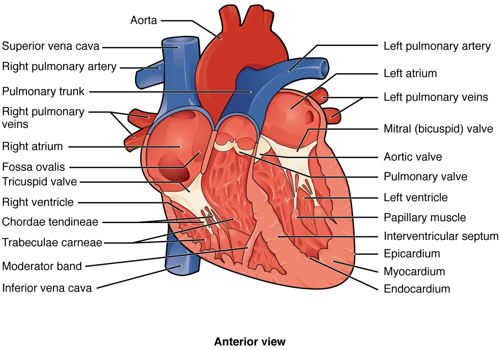
The circulatory system exists to maintain adequate perfusion to all tissues. Perfusion means oxygen, glucose, and key nutrients are delivered to cells in sufficient amounts while metabolic waste products are removed and excreted. When perfusion drops, cells shift from normal aerobic metabolism to less efficient pathways, acid builds, and organ systems begin to fail.
In addition to perfusion, the cardiovascular system supports homeostasis by helping regulate fluid balance, acid-base balance, and temperature. It also distributes immune system components and circulates clotting factors to help stop bleeding. In EMS, many “big” emergencies are fundamentally perfusion problems.
The Perfusion Triangle: Pump, Container, Fluid
A useful framework from the lecture is the perfusion triangle. Adequate perfusion depends on three components working together. The heart is the pump. The blood vessels are the container. The blood is the fluid that fills the system.
If any one part fails, the patient is at risk for shock, defined as inadequate tissue perfusion. This model helps EMTs structure their thinking during assessment. Is the pump failing, is the container too large or leaking, or is the fluid volume inadequate or unable to carry oxygen effectively.
Blood Flow Through the Heart: A Practical Map
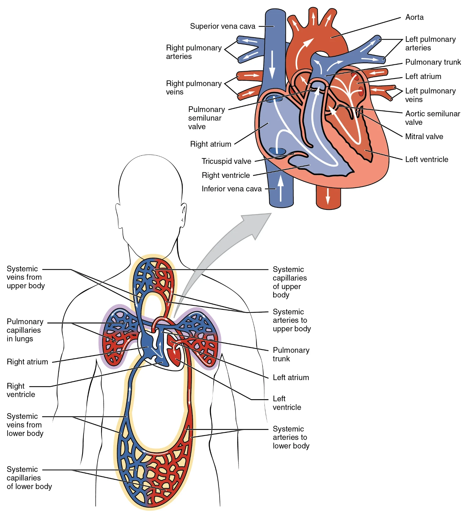
Deoxygenated blood returns to the heart from the body through the venae cavae. The superior vena cava carries blood from the upper body, and the inferior vena cava carries blood from the lower body. This blood enters the right atrium. When the right atrium contracts, blood passes through the tricuspid valve into the right ventricle.
When the right ventricle contracts, blood is pushed through the pulmonary valve into the pulmonary artery and sent to the lungs. In the pulmonary capillaries, carbon dioxide is removed and oxygen is loaded. Oxygenated blood returns to the heart through the pulmonary veins into the left atrium. From the left atrium, blood flows through the bicuspid valve into the left ventricle.
The left ventricle is the strongest chamber because it pumps blood through the aortic valve into the aorta and out to the systemic circulation. This is why left-sided pump failure can produce rapid respiratory distress and pulmonary edema, and why left ventricular performance is central to perfusion.
Two Circuits, One Double Pump
The heart functions as a double pump. The right side drives the pulmonary circuit, sending deoxygenated blood to the lungs. The left side drives the systemic circuit, delivering oxygenated blood to the body. The two circuits are connected in series, meaning failure of one side can affect the other.
For EMTs, this explains why a patient with poor ventilation can become hypoxic and unstable quickly and why a patient with severe left-sided failure can develop respiratory symptoms even though the primary issue is cardiac.
Valves, Systole, and Diastole
Valves maintain one-way blood flow. The atrioventricular valves sit between atria and ventricles. The tricuspid valve has three cusps on the right side. The bicuspid valve has two cusps on the left side and is also called the mitral valve. The semilunar valves include the pulmonary valve and aortic valve.
When ventricles contract, this is systole, and arterial pressures reach their peak. When ventricles relax and fill, this is diastole. These phases matter clinically because coronary arteries primarily fill during diastole, meaning very fast heart rates can reduce coronary perfusion and contribute to ischemia.
Electrical Cells and Mechanical Cells
Heart muscle includes electrical cells and mechanical cells. Electrical cells generate and conduct impulses. Mechanical cells contract in response, producing the pumping action. This separation helps explain how dysrhythmias can reduce perfusion even when the heart is “still beating.” If electrical conduction is disorganized or too slow or too fast, mechanical pumping becomes ineffective.
Blood Vessels: Arteries, Veins, and Capillaries
Blood vessels form the container portion of the perfusion triangle. The lecture emphasizes a key rule. Arteries carry blood away from the heart. Veins return blood to the heart. Capillaries are where exchange occurs.
Blood leaves the left ventricle into the aorta, then travels through progressively smaller arteries and arterioles. Arterioles connect to capillary beds where oxygen is delivered and waste is collected. Blood then flows into venules, then veins, returning to the venae cavae and right atrium.
In capillary beds, precapillary sphincters regulate flow. This allows the body to redirect blood to tissues that need it most, such as working muscles or vital organs during stress. In shock, this redistribution becomes a core compensatory mechanism, and as it fails, perfusion collapses.
Pressure Differences and Venous Valves
Arterial blood is under higher pressure than venous blood. This helps drive forward flow. Because venous pressures are lower, the venous system includes one-way valves that support return flow back to the heart, especially from the extremities. From an EMS perspective, this reinforces why venous return is sensitive to volume status and why position and muscle activity can affect perfusion.
Blood: Plasma and Formed Elements
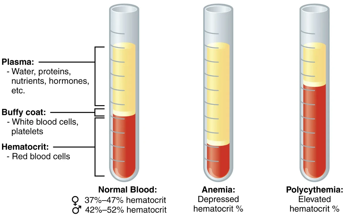
Blood is the fluid portion of the perfusion triangle. It consists of plasma, the fluid medium, and formed elements, the cellular components carried within the plasma. Formed elements include red blood cells, white blood cells, and platelets.
Red blood cells contain hemoglobin, which carries oxygen. White blood cells support immune defense. Platelets contribute to clotting. From an EMS perspective, anemia, bleeding, or hemoglobin dysfunction are “fluid problems” because oxygen delivery decreases even if blood pressure appears normal initially.
Average adult blood volume is approximately five liters, but actual volume varies by size and body composition. Loss of circulating volume compromises preload and can rapidly reduce cardiac output.
Cardiac Output: The Core Perfusion Equation
Cardiac output is the volume of blood pumped by the ventricles in one minute. The lecture defines it as heart rate multiplied by stroke volume. Stroke volume is the amount pumped with each contraction.
This relationship helps EMTs interpret clinical findings. A patient can maintain cardiac output with an increased heart rate even as stroke volume falls, but this compensation has limits. When compensation fails, perfusion drops, mental status changes, skin signs worsen, and blood pressure may fall late in the process.
Stroke Volume Drivers: Preload, Contractility, and Afterload
Stroke volume depends on the difference between end diastolic volume and end systolic volume. End diastolic volume represents how much blood is in the ventricle just before contraction. End systolic volume is what remains after contraction.
Three primary factors determine stroke volume. Preload is the stretch on the ventricle prior to contraction and reflects venous return and filling. Contractility is the strength of contraction. Afterload is the resistance the ventricle must overcome to eject blood, which is influenced by systemic vascular resistance.
Clinically, dehydration and hemorrhage reduce preload. Cardiac ischemia can reduce contractility. Vasoconstriction and hypertension increase afterload. All three can reduce effective perfusion and contribute to shock.
Frank-Starling Mechanism
A unique property of cardiac muscle is that increased stretch leads to stronger contraction, within physiologic limits. This is the Frank-Starling mechanism, also called Starling’s law. As preload increases, the ventricle contracts more forcefully, increasing stroke volume.
The lecture uses a balloon analogy. More filling creates more force when released. In EMS terms, adequate volume status supports stroke volume, but excessive preload in a failing heart can worsen congestion and respiratory distress. This is why careful assessment is essential when considering fluid administration under protocol.
Blood Types, Transfusion Compatibility, and Hemolytic Reactions
Blood typing is based on antigens on red blood cells and antibodies in plasma. Incompatible transfusions can trigger a hemolytic transfusion reaction, where the immune system destroys transfused red blood cells. This is life-threatening and is a core reason blood typing and compatibility checks exist.
ABO typing includes A, B, AB, and O. Type A has A antigens and anti-B antibodies. Type B has B antigens and anti-A antibodies. Type AB has both antigens and no anti-A or anti-B antibodies, making AB the universal recipient for ABO typing. Type O has no A or B antigens but has both antibodies, making O the universal donor for ABO typing.
Rh Factor and Why It Matters
The Rh system involves the presence or absence of the Rh antigen. Rh positive individuals have the antigen. Rh negative individuals do not and can form antibodies if exposed to Rh positive blood. A second exposure can cause severe hemolysis.
Rh status also has clinical relevance in pregnancy when maternal and fetal Rh types differ. For EMT education, the practical takeaway is that Rh compatibility matters in transfusion and maternal-fetal considerations, and it should be documented accurately when known.
The Cardiac Conduction System
The lecture outlines the conduction system components. The sinoatrial node is the primary pacemaker and typically fires at 60 to 100 beats per minute. Impulses spread through the atria and reach the atrioventricular node. A delay at the AV node allows ventricular filling before the impulse travels down the bundle of His, through right and left bundle branches, and into the Purkinje fibers to depolarize ventricles.
This electrical pathway is fundamental to understanding ECG interpretation and dysrhythmias. Even at the EMT level, recognizing that electrical problems cause mechanical perfusion problems supports better assessment and earlier escalation when patients deteriorate.
Coronary Arteries and Diastolic Filling
The myocardium requires continuous oxygen and nutrient delivery because the heart does not rest. Coronary arteries branch from the aorta just above the aortic valve and are the first vessels to receive oxygenated blood after left ventricular ejection.
A critical point from the lecture is that coronary arteries fill primarily during diastole because they are compressed during systole. This helps explain why very high heart rates reduce coronary perfusion time and can worsen ischemia, chest pain, or pump failure.
Critical Thinking Questions
A patient is tachycardic with cool, clammy skin but has a normal blood pressure. Using the perfusion triangle, how would you evaluate whether the primary problem is pump failure, container failure, or fluid loss?
A patient has a very rapid heart rate and chest discomfort. How might reduced diastolic time affect coronary perfusion and contribute to worsening symptoms?
A trauma patient has significant bleeding and becomes increasingly anxious and pale. How do preload and stroke volume change as blood volume falls, and what compensations might you expect to see early?
A patient with suspected heart failure is short of breath with crackles and edema. How can the Frank-Starling mechanism explain why increased preload can worsen respiratory symptoms in a failing heart?
A patient reports a history of “valve problems.” How would you use heart anatomy, valve function, and systole versus diastole concepts to guide your focused history and assessment priorities?
Image Credits: Image from Anatomy and Physiology by OpenStax, licensed under CC BY 4.0