
Basic Anatomy and Physiology: Genitourinary System
Prehospital Academy Lesson 7.5
Why the Urinary System Matters in EMS
The urinary system is a major homeostasis regulator. It controls fluid balance, filters metabolic waste, and helps stabilize electrolyte concentration and pH. When these processes fail, patients can present with weakness, dysrhythmias, altered mental status, respiratory compensation, or shock.
The kidneys also participate in vitamin D activation and play a long-term role in blood pressure regulation. For prehospital care, it is often helpful to think of the kidneys as a “control center” for volume, electrolytes, and acid-base balance that influences nearly every body system.
Core Functions of the Kidneys
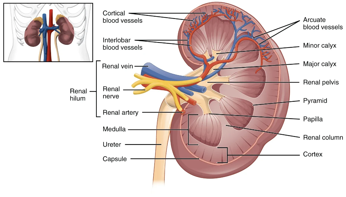
The kidneys filter water and waste products from blood to form urine. They conserve fluid when the patient is dehydrated and allow increased excretion of water when the patient has excess volume. This is one reason dehydration can progress quickly in vulnerable patients, including older adults, febrile patients, and those with vomiting or diarrhea.
The kidneys help regulate acid-base balance by controlling bicarbonate reabsorption and hydrogen ion secretion. They also work with the cardiovascular system to regulate blood pressure. In addition, the kidneys produce most erythropoietin, which stimulates red blood cell production, linking renal function to long-term oxygen-carrying capacity.
Urinary System Anatomy: The Flow Path
The urinary system includes two kidneys, two ureters, the urinary bladder, and the urethra. After urine forms in the kidneys, it travels down the ureters to the bladder for storage and exits the body through the urethra. Urination may also be referred to as voiding or micturition.
The bladder’s approximate storage capacity is around 500 mL in women and 700 mL in men, but the urge to urinate is often felt at lower volumes, typically around 200 to 350 mL. In EMS, urinary retention, reduced output, or dark concentrated urine can provide important clues about hydration status and perfusion.
Renal Blood Supply and Why It Is Significant
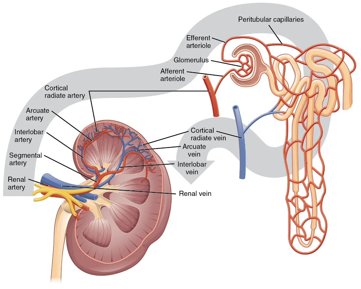
Kidneys require substantial blood flow to perform filtration. Blood enters each kidney through the renal artery and exits via the renal vein. This high perfusion demand is why shock can rapidly impair kidney function and why reduced urine output can be an early clue of poor systemic perfusion.
From a systems perspective, the body’s approximate five liters of blood pass through the kidneys repeatedly throughout the day. The lecture emphasizes the magnitude of renal filtration and why even small changes in renal function can meaningfully disrupt homeostasis.
Nephrons: The Functional Units
Each kidney contains approximately one million nephrons. Each nephron includes a filter (the glomerulus) and a tubule system that modifies the filtered fluid. The nephron works through a two-step concept. First, the glomerulus filters the blood. Second, the tubules reabsorb needed substances and allow wastes and excess ions to remain in the filtrate, which becomes urine.
This “filter then fine-tune” concept is one of the most important takeaways for EMT physiology. Most of what gets filtered is not ultimately excreted. The kidney intentionally returns key substances to the bloodstream and selectively removes what the body needs to eliminate.
From Filtrate to Urine: Reabsorption and Concentration
The kidney initially produces a large volume of primary urine (glomerular filtrate), which is then dramatically reduced through reabsorption to a much smaller daily urine output. As filtrate flows through nephron tubules and collecting ducts, the kidneys reabsorb essential substances such as water, glucose, and electrolytes back into the blood while leaving wastes behind.
This process is why hydration status changes urine appearance. Well-hydrated urine is typically clear to pale yellow. Dehydration produces darker, more concentrated urine that may have a mild ammonia odor. In the field, these observations are supportive findings that should be paired with vital signs, mental status, skin findings, and history.
Renal Corpuscle: Glomerulus and Bowman’s Capsule
The renal corpuscle consists of the glomerulus, a tangled capillary network, and Bowman’s capsule, a cup-shaped structure surrounding the glomerulus. Bowman’s capsule captures the filtrate and directs it into the proximal convoluted tubule. This is where urine formation begins, and it sets the stage for the selective reabsorption and secretion that follow.
Proximal Convoluted Tubule: High-Volume Reabsorption
The proximal convoluted tubule performs significant reabsorption of water and solutes. Its lining contains microvilli, which increase surface area and improve reabsorption efficiency. This is where critical substances such as sodium, chloride, and glucose are reclaimed to maintain plasma composition and preserve energy resources.
Loop of Henle: Concentrating Mechanism
The loop of Henle creates a concentration gradient in the surrounding renal medulla, enabling the kidney to concentrate urine and regulate water balance. By controlling water and electrolyte movement, the loop supports the body’s ability to conserve water when dehydrated and adjust urine concentration based on physiologic need.
For EMT-level understanding, the key point is that the kidney can intentionally “save water” and still excrete wastes, but only if this concentrating mechanism is functioning properly.
Distal Convoluted Tubule: Fine Tuning and pH Regulation
The distal convoluted tubule continues electrolyte fine-tuning, particularly sodium and calcium handling, and contributes to urine pH regulation. It does this by secreting hydrogen ions and reabsorbing bicarbonate ions, allowing the body to adjust acid-base balance over time.
This helps explain why kidney dysfunction can contribute to metabolic acidosis and why chronically ill patients with renal disease may show compensatory respiratory patterns.
Collecting Ducts: Final Adjustment Based on Hydration Status
Collecting ducts receive filtrate from multiple nephrons and adjust final urine concentration. They selectively reabsorb water and help regulate sodium, potassium, and hydrogen ion balance. This is a major point where hormones influence how much water is conserved versus excreted.
RAAS: Renin-Angiotensin-Aldosterone System
The renin-angiotensin-aldosterone system regulates blood pressure and blood volume over the long term. When blood pressure falls, kidneys release renin. Renin acts on angiotensinogen from the liver to form angiotensin I. Angiotensin-converting enzyme converts angiotensin I to angiotensin II, the active hormone.
Angiotensin II causes arteriolar vasoconstriction, increasing blood pressure. It also stimulates the adrenal glands to release aldosterone and the pituitary to release antidiuretic hormone. Aldosterone promotes sodium reabsorption and potassium excretion. ADH promotes water retention. The net effect is increased blood volume and increased blood pressure.
For EMTs, the takeaway is that the kidneys are not passive filters. They actively participate in blood pressure control through hormonal pathways that alter vascular tone and fluid retention.
Electrolyte Regulation: Sodium, Potassium, and Others
Electrolytes such as sodium, potassium, chloride, bicarbonate, calcium, and phosphate are essential for neurologic function, muscle contraction, fluid balance, and cardiac conduction. The kidneys maintain electrolyte balance by filtering them and then selectively reabsorbing or excreting them based on the body’s needs.
A practical example is aldosterone’s influence on sodium reabsorption. When sodium levels are low or when RAAS is activated, kidneys retain more sodium, which drives water retention. Potassium levels are also tightly regulated, and abnormal potassium can lead to dangerous dysrhythmias. While EMTs do not correct electrolyte abnormalities definitively, recognizing their clinical consequences supports early identification of unstable patients.
Acid-Base Balance: Bicarbonate and Hydrogen Ions
The kidneys maintain pH using two key mechanisms. They reabsorb bicarbonate back into the bloodstream and they secrete hydrogen ions into the urine. By altering how much bicarbonate is conserved or excreted and how many hydrogen ions are secreted, the kidneys help maintain normal blood pH.
When blood becomes too acidic, kidneys excrete more hydrogen ions and retain bicarbonate, raising pH toward normal. When blood becomes too alkaline, kidneys can excrete bicarbonate and conserve hydrogen ions, lowering pH toward normal. This supports the body’s normal pH range of 7.35 to 7.45, which is necessary for enzymes and physiologic processes to function effectively.
Clinical Reinforcement for EMTs
In the field, renal physiology shows up in many subtle ways. Dehydration reduces perfusion and can lower urine output. Renal dysfunction can worsen acid-base imbalance and contribute to altered mental status. RAAS activation is part of the body’s attempt to preserve perfusion during volume loss, but compensation has limits.
When evaluating a patient with weakness, confusion, dysrhythmias, or signs of dehydration, consider the possibility of renal or electrolyte involvement, especially in older adults, patients with diabetes, patients on diuretics, or those with chronic kidney disease.
Critical Thinking Questions
A patient with vomiting and diarrhea has dark, concentrated urine and tachycardia. How does decreased circulating volume influence renal perfusion and RAAS activation, and what assessment findings would support dehydration severity?
A dialysis patient is weak and bradycardic with an irregular pulse. How does renal regulation of potassium relate to the risk of dysrhythmia, and what information would you prioritize in your history and handoff?
A patient with suspected sepsis has low blood pressure and reduced urine output. How does the perfusion triangle concept help you explain why kidney function is often affected early in shock states?
A patient is breathing rapidly and deeply and has a history of renal disease. How can kidney involvement contribute to acid-base imbalance, and what clinical cues might suggest metabolic acidosis with respiratory compensation?
A hypertensive patient takes multiple blood pressure medications and becomes dizzy when standing. How could changes in vascular tone, fluid balance, and renal regulation contribute to orthostatic symptoms, and what vital sign trends would you track?
Image Credits: Image from Anatomy and Physiology by OpenStax, licensed under CC BY 4.0