
Basic Anatomy and Physiology: Pulmonary System
Prehospital Academy Lesson 7.4
What the Respiratory System Does for the Patient
The primary role of the respiratory system is to move air in and out of the body and exchange carbon dioxide waste for oxygen. It also filters particulates, traps microorganisms in mucus, and helps remove them. Beyond gas exchange, the respiratory system plays a major role in regulating pH by controlling carbon dioxide removal. It enables speech and contributes to the sense of smell.
For EMS, this is why respiratory emergencies are not just “breathing problems.” They can become acid-base problems, perfusion problems, neurologic problems, and rapidly escalating airway problems.
Key Terms EMTs Must Not Confuse
Ventilation is the physical movement of air in and out of the lungs. It includes inhalation and exhalation. Oxygenation is the process of loading oxygen onto hemoglobin in red blood cells. Respiration is the exchange of oxygen and carbon dioxide, which occurs both externally in the lungs and internally at the tissues.
These distinctions matter clinically. A patient can ventilate but not oxygenate well. A patient can oxygenate but not ventilate effectively. A patient can move air and have adequate oxygen saturation while still failing at cellular respiration due to shock or toxin exposure.
Upper Airway Anatomy and Protective Functions
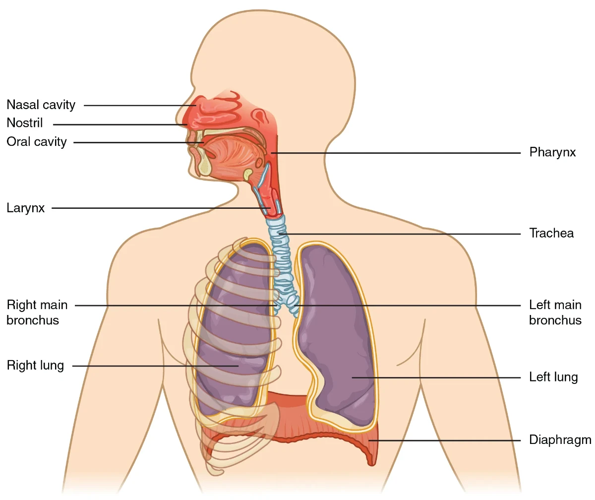
The upper airway warms, filters, and humidifies incoming air. Air entering through the nose passes the nasal cavity into the nasopharynx. Air entering through the mouth passes the oral cavity into the oropharynx, which is a shared passage for air and food. The posterior oropharynx has sensory innervation that can trigger the gag reflex, providing protection against aspiration.
The hard palate and soft palate separate the oral and nasal cavities. The laryngopharynx lies below the oropharynx and extends from the epiglottis toward the entrance of the trachea. Because food and air share this region, the epiglottis plays a critical protective role by closing during swallowing and diverting food into the esophagus.
Lower Airway Anatomy: From Trachea to Alveoli
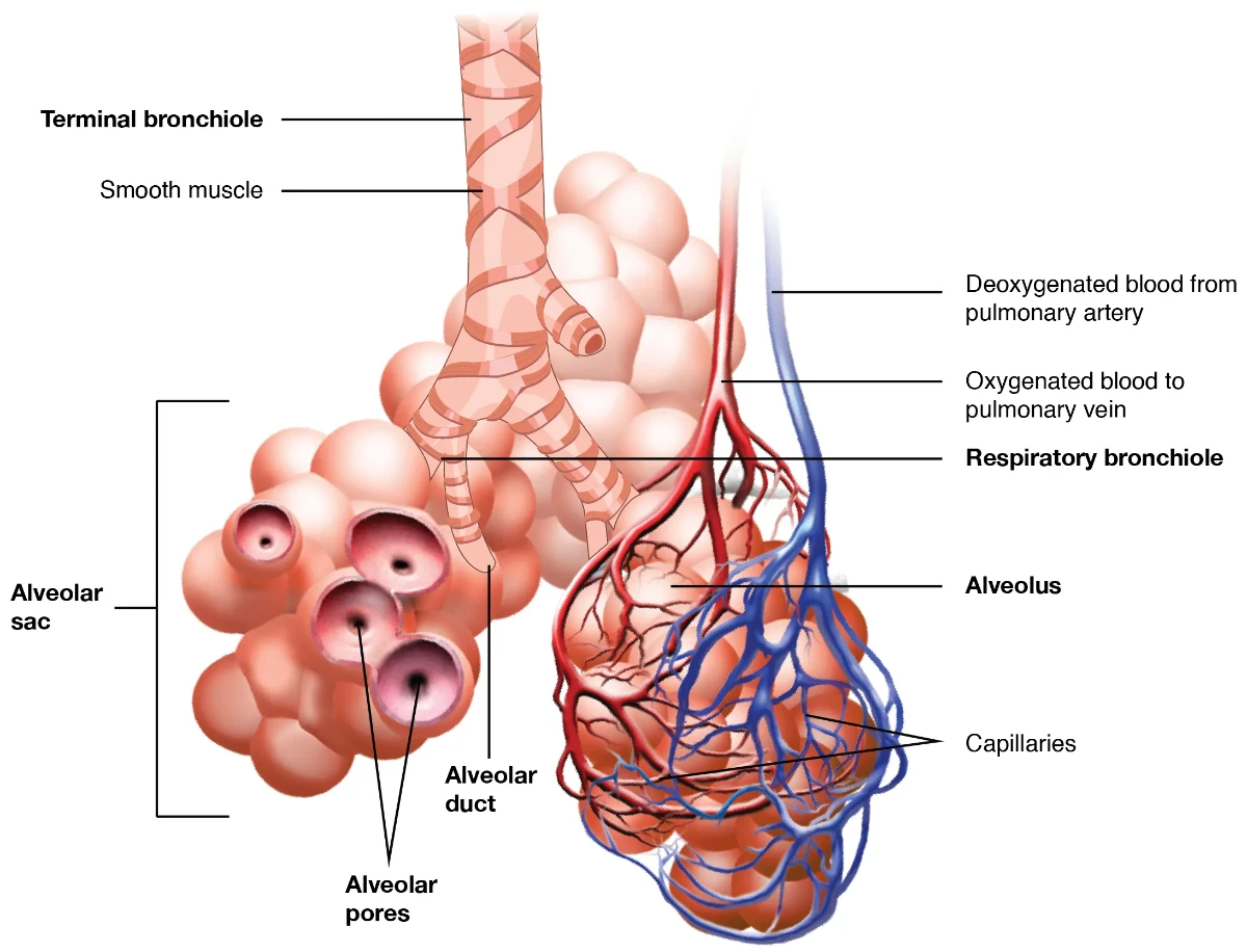
The larynx connects the pharynx to the trachea, supports air passage, helps prevent solids and liquids from entering the lower airway, and contains the vocal cords. Air travels down the trachea to the carina, where the trachea divides into the right and left mainstem bronchi. These divide into smaller bronchi, then bronchioles, then alveolar ducts that lead to the alveoli.
The alveoli are the functional units where gas exchange occurs. They are often described as grape-like clusters, with each small sac supporting diffusion between air and pulmonary capillary blood.
Alveolar Gas Exchange: External vs Internal Respiration
Each alveolus has a thin wall, approximately one cell layer thick, allowing diffusion to occur efficiently. Carbon dioxide diffuses out of pulmonary capillary blood into the alveolus to be exhaled. Oxygen diffuses from the alveolus into the blood and binds to hemoglobin on red blood cells. This is external respiration because the exchange occurs with the external environment.
Internal respiration occurs at the tissue level, where oxygen leaves the blood and enters cells while carbon dioxide enters the blood to be transported back to the lungs. In EMS, inadequate perfusion can disrupt internal respiration even when lung function appears adequate.
Surfactant, Compliance, and Atelectasis
Alveoli contain pulmonary surfactant, which reduces surface tension. This improves pulmonary compliance and helps prevent atelectasis, or alveolar collapse. Clinically, reduced compliance increases the work of breathing and can contribute to respiratory fatigue, especially when the patient must generate higher pressures to ventilate.
How Oxygen Is Transported in Blood
Only a small amount of oxygen is dissolved directly in plasma. Most oxygen is carried bound to hemoglobin within red blood cells. Each hemoglobin molecule can carry four oxygen molecules, and oxygenation describes the loading of oxygen onto these binding sites.
This is why hemoglobin matters. Even with adequate ventilation, oxygen delivery can be compromised by low hemoglobin, carbon monoxide exposure, or abnormal hemoglobin function.
How Carbon Dioxide Is Transported in Blood
Carbon dioxide is transported in three forms. A portion is dissolved directly in plasma. Most is transported as bicarbonate in plasma. A smaller portion is transported bound to hemoglobin within red blood cells.
This matters because CO2 is tightly connected to pH. When CO2 rises, acidity increases. When CO2 falls, pH rises. EMS ventilation support is often about controlling CO2 as much as it is about delivering oxygen.
Breathing Mechanics and Boyle’s Law
Ventilation is driven by pressure gradients governed by Boyle’s law, which states that gas pressure is inversely proportional to volume. When thoracic volume increases, intrathoracic pressure decreases. Air moves from higher pressure to lower pressure, so atmospheric air flows into the lungs during inhalation.
During inspiration, the diaphragm contracts and flattens, and intercostal muscles elevate and expand the rib cage. This increases thoracic volume and decreases intrathoracic pressure. During exhalation, these muscles relax, volume decreases, pressure rises, and air flows out. Inhalation is active and requires energy. Exhalation is typically passive.
Lung Volumes EMTs Should Recognize
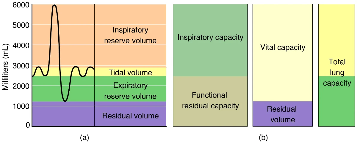
Tidal volume is the amount of air moved during a normal breath, commonly around 500 mL in an adult. Inspiratory reserve volume is additional air that can be inhaled beyond a normal breath. Expiratory reserve volume is additional air that can be forcefully exhaled after a normal exhalation. Residual volume remains in the lungs after maximal exhalation and helps keep alveoli open.
In the field, you do not measure these volumes directly, but they explain important patterns. Shallow breathing reduces effective alveolar ventilation. Fatigue reduces inspiratory capacity. Trapped air and poor exhalation can worsen work of breathing and CO2 retention in obstructive disease.
Dead Space: Why Bag-Valve Mask Technique Matters
Anatomic dead space is air in the respiratory passages that does not participate in gas exchange. This includes the upper airway and conducting airways down to the terminal bronchioles. A helpful approximation is about 1 mL per pound of body weight.
This concept explains why effective BVM ventilation requires adequate tidal volumes. If you ventilate with too little volume, you primarily move air in and out of dead space without delivering meaningful gas exchange at the alveoli. Physiologic dead space includes alveoli that cannot participate in exchange due to collapse or damage.
Gas Laws and Partial Pressures
Gases exert pressure, and in a mixture each gas contributes its own partial pressure. Dalton’s law states total pressure is the sum of the partial pressures of individual gases. Henry’s law states the concentration of a gas dissolved in a liquid depends on the gas’s solubility and its partial pressure.
Diffusion occurs when gases move from areas of higher partial pressure to lower partial pressure until equilibrium is approached. The greater the gradient, the faster diffusion occurs. These principles explain why oxygen moves from alveoli into blood and why carbon dioxide moves from blood into alveoli.
Why Oxygen and CO2 Move: External Respiration as Diffusion
Inspired air has a high partial pressure of oxygen. In the alveoli, oxygen partial pressure is lower than in the atmosphere but still higher than in pulmonary arterial blood. This gradient drives oxygen diffusion into the blood until equilibrium is reached.
For carbon dioxide, the gradient is reversed. Pulmonary arterial blood has a higher partial pressure of CO2 than alveolar air, so CO2 diffuses into the alveoli to be exhaled. In EMS terms, anything that reduces alveolar ventilation, reduces diffusion surface area, or thickens the membrane can impair gas exchange.
Control of Breathing: Sensors, Control Center, Effectors
Involuntary breathing is regulated by a control system with three parts. Sensors detect CO2, oxygen, and pH changes. The control center in the brainstem coordinates a response. Effectors are the respiratory muscles that change rate and depth of breathing.
The respiratory control center includes the dorsal and ventral respiratory groups in the medulla and the pontine respiratory group in the pons. These groups create rhythmic breathing and adjust ventilation based on physiologic needs.
CO2, pH, and Ventilation
CO2 is a powerful driver of ventilation. When CO2 rises, pH falls, and chemoreceptors respond by increasing the rate and depth of breathing to exhale more CO2. This helps reduce hydrogen ion concentration and restore pH balance. When CO2 falls and pH rises, ventilation decreases to allow CO2 levels to normalize.
This is why hypoventilation can be dangerous even when oxygen saturation initially appears acceptable. CO2 retention can drive acidosis, altered mental status, and eventual respiratory failure.
Hypoxic Drive: What It Is and What It Is Not
In some chronic CO2 retainers, such as certain COPD patients, the stimulus to breathe may shift from CO2-driven control to oxygen-driven control. This is described as hypoxic drive. The practical EMS takeaway is not to withhold oxygen, but to titrate oxygen appropriately and monitor ventilation status closely. Oxygenation and ventilation are different problems, and CO2 retention must be assessed and managed within protocol.
Clinical Reinforcement for EMTs
Use respiratory anatomy and physiology to guide a structured assessment. Confirm airway patency, evaluate the work of breathing, assess ventilation effectiveness, and then address oxygenation. Remember that oxygen saturation is a snapshot of oxygenation, not ventilation. A patient can have rising CO2 and worsening acidosis while SpO2 looks acceptable.
BVM effectiveness depends on sufficient tidal volume to overcome dead space and reach the alveoli. Inadequate volumes lead to ineffective ventilation and rapid deterioration. Correct technique, seal, and rate matter as much as oxygen delivery.
Critical Thinking Questions
A patient is breathing rapidly but shallowly and has normal SpO2. How does anatomic dead space explain why this patient may still have inadequate alveolar ventilation and rising CO2?
A patient with suspected COPD is lethargic with slow breathing and wheezing. How do CO2, pH, and hypoxic drive concepts guide your priorities for ventilation support and oxygen titration?
A patient has pneumonia and increasing work of breathing. How could thickened alveolar membranes and reduced effective surface area impair diffusion even if ventilation rate increases?
During BVM ventilation, you notice minimal chest rise and poor compliance. How might surfactant function, atelectasis, or decreased lung compliance affect your ventilation strategy?
A trauma patient is pale and anxious with adequate breath sounds and acceptable SpO2. How can poor perfusion impair internal respiration and oxygen delivery to tissues despite adequate external respiration?
Image Credits: Image from Anatomy and Physiology by OpenStax, licensed under CC BY 4.0