
Basic Anatomy and Physiology: Neurological System
Prehospital Academy Lesson 7.2
The Cellular Foundation: The Neuron and Synaptic Transmission
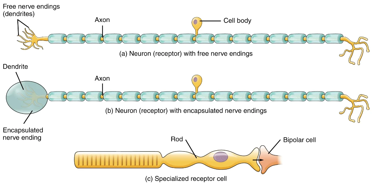
At the microscopic level, the nervous system is composed of billions of specialized cells known as neurons. These cells are unique in their ability to conduct electrical impulses and communicate via chemical messengers. A neuron consists of three primary structures:
Dendrites: Branch-like projections that receive incoming signals from other neurons.
Cell Body (Soma): The metabolic heart of the cell, containing the nucleus and responsible for protein synthesis and energy production.
Axon: A long, slender projection that conducts electrical impulses (action potentials) away from the cell body toward a target tissue or another neuron.
Communication between neurons occurs at the synapse, a microscopic gap where the electrical signal is converted into a chemical one. When an impulse reaches the axon terminal, it triggers the release of neurotransmitters. These molecules cross the synaptic cleft and bind to receptors on the target cell, either exciting or inhibiting further electrical activity. In the EMS context, many pharmacological interventions (such as Albuterol or Epinephrine) work by mimicking or blocking these naturally occurring neurotransmitters.
Anatomical Divisions: Central and Peripheral Systems
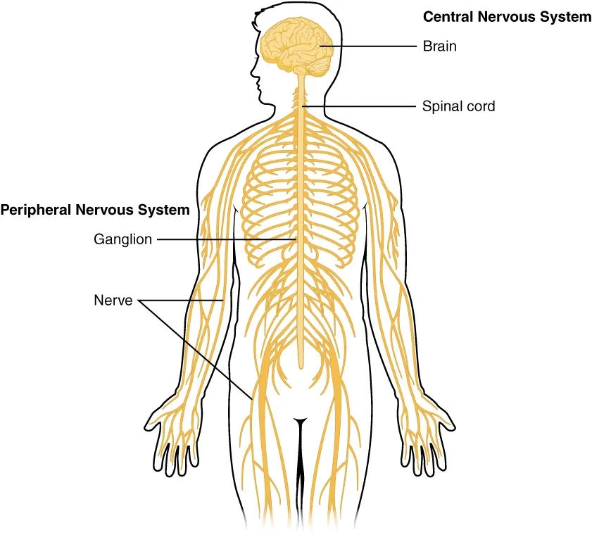
The human nervous system is divided into two major anatomical branches: the Central Nervous System (CNS) and the Peripheral Nervous System (PNS).
The Central Nervous System (CNS)
The CNS consists of the brain and the spinal cord, encased within the protective structures of the skull and vertebral column. The brain is further divided into:
The Cerebrum: The largest part of the brain, responsible for high-level functions. It is divided into four lobes: the Frontal (executive function and motor control), Parietal (sensory processing), Temporal (hearing and memory), and Occipital (vision).
The Cerebellum: Located posteriorly, it coordinates balance, posture, and complex muscle movements.
The Brain Stem: The most primitive and vital part of the brain, comprising the midbrain, pons, and medulla oblongata. It regulates life-sustaining functions such as respiratory rate, heart rate, and blood pressure.
The Peripheral Nervous System (PNS)
The PNS acts as the "wiring" that connects the CNS to the rest of the body. It includes 12 pairs of cranial nerves—which primarily manage sensory and motor functions of the head and neck (with the exception of the Vagus nerve)—and 31 pairs of spinal nerves that exit the spinal cord to innervate the trunk and limbs.
The Autonomic Engine: Balance and Stress Response
The motor division of the PNS is further subdivided into the somatic (voluntary) and autonomic (involuntary) systems. The Autonomic Nervous System (ANS) is of particular interest to EMS providers as it governs the "fight-or-flight" and "rest-and-digest" responses.
The Sympathetic Nervous System prepares the body for stress. Activation leads to bronchodilation, tachycardia, pupil dilation, and the shunting of blood from the GI tract to the skeletal muscles and brain. Conversely, the Parasympathetic Nervous System promotes energy conservation, slowing the heart rate and increasing digestive activity. Understanding this balance is critical when assessing patients in shock or those who have ingested toxins that mimic or inhibit these systems (e.g., organophosphate poisoning).
Sensory Perception and the Pathophysiology of Pain
Pain is a protective mechanism signaled by nociceptors. EMS providers must distinguish between different types of pain to narrow their differential diagnosis:
Somatic Pain: Originates from the skin, muscles, or bones. It is typically sharp and well-localized.
Visceral Pain: Arises from internal organs. Because organ innervation is less precise, this pain is often described as dull, aching, or "pressure," and is frequently poorly localized.
Referred Pain: Pain felt in an area distant from the actual source. A classic example is Kehr’s Sign (left shoulder pain) resulting from splenic rupture, or jaw pain during a myocardial infarction.
Neuropathic Pain: Caused by direct damage to the nerves themselves, often described as "burning" or "electric shocks."
Cerebral Hemodynamics and the Monroe-Kellie Doctrine
To maintain consciousness and cellular health, the brain requires constant perfusion. This is governed by Cerebral Perfusion Pressure (CPP), which is the pressure gradient that drives blood into the brain.
The mathematical relationship is: CPP = MAP - ICP.
Mean Arterial Pressure (MAP): The average pressure in the systemic arteries. It is calculated as: Diastolic BP + 1/3(Pulse Pressure).
Intracranial Pressure (ICP): The pressure exerted by the brain tissue, blood, and cerebrospinal fluid (CSF) inside the rigid skull.
The Monroe-Kellie Doctrine states that the skull is a rigid container with a fixed volume. If one component (like blood from a hemorrhage) increases, another (like CSF) must decrease to maintain a stable ICP. When compensatory mechanisms fail, ICP rises, and CPP drops, leading to brain ischemia. Clinically, this may manifest as Cushing’s Triad: hypertension (widening pulse pressure), bradycardia, and irregular respirations—a late and ominous sign of brain herniation.
Critical Thinking Questions
How does the "Monroe-Kellie Doctrine" explain why a patient with a slow intracranial bleed might initially appear neurologically intact before suddenly deteriorating?
A patient presents with a heart rate of 40, a BP of 190/70, and irregular breathing. Based on your understanding of CPP and ICP, what is the physiological reason for the bradycardia?
Explain why visceral pain from an abdominal organ like the appendix might be felt globally across the abdomen before localizing to the Right Lower Quadrant.
Using the concept of neurotransmission, why would a drug that blocks Acetylcholine receptors (an anticholinergic) result in tachycardia and dry skin?
How might an injury to the temporal lobe specifically affect an EMT’s ability to obtain a patient history, even if the patient's motor function is intact?
Image Credits: Image from Anatomy and Physiology by OpenStax, licensed under CC BY 4.0