
Basic Anatomy and Physiology: Integumentary and Musculoskeletal Systems
Prehospital Academy Lesson 7.1
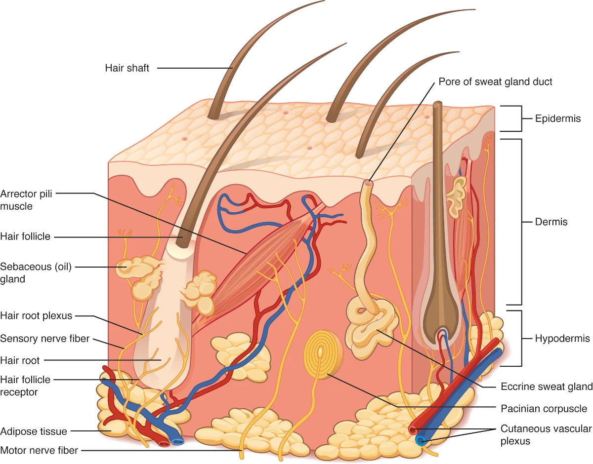
The Integumentary System: The Body's First Line of Defense
The integumentary system, consisting of the skin, hair, nails, and associated glands, is the body's largest organ and serves as its primary protective barrier. For an EMT, the skin provides immediate and valuable clues to a patient's overall health and perfusion status. Its functions are diverse and critical for survival.
Anatomy and Key Functions:
Protection: The skin acts as a formidable shield against pathogens, harmful chemicals, and ultraviolet radiation. The outermost layer, the epidermis, is constantly renewing itself, providing a durable and resilient barrier. Any breach in this barrier, such as a laceration or burn, significantly increases the risk of infection.
Temperature Regulation: The skin plays a vital role in thermoregulation. Blood vessels within the dermis can dilate to release heat or constrict to conserve it. Sweat glands produce perspiration, which cools the body through evaporation. The failure of this system can lead to life-threatening conditions like heat stroke.
Sensation: Richly supplied with nerve endings, the skin is a complex sensory organ, detecting touch, pressure, pain, and temperature. This function is critical for a patient's ability to react to their environment and for an EMT's assessment of neurological function.
Water Balance and Vitamin D Synthesis: The skin forms a waterproof seal that prevents dehydration. It is also the site of Vitamin D synthesis, a crucial process initiated by exposure to sunlight.
Clinical Significance for EMTs:
An EMT's assessment of the skin can reveal a wealth of information. Skin color (e.g., paleness, cyanosis, flushing), temperature, and moisture level are key indicators of a patient's circulatory status and can signal conditions like shock or fever. The integumentary system is also the most common site of injury in trauma patients, and EMTs must be proficient in managing a wide range of soft-tissue injuries, from simple abrasions to complex avulsions and burns, while always prioritizing bleeding control and infection prevention.
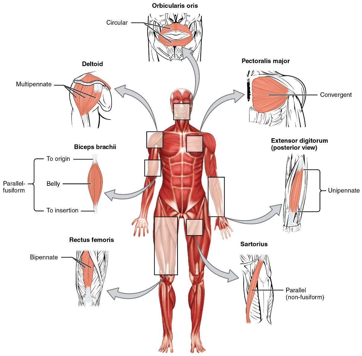
The Musculoskeletal System: The Framework for Movement and Protection
The musculoskeletal system, a complex network of bones, muscles, joints, ligaments, and tendons, provides the body with its structure, enables movement, and protects vital internal organs. For EMTs, a solid understanding of this system is essential for assessing and managing the vast majority of traumatic injuries.
Anatomy and Key Functions:
The Skeletal System: Comprising 206 bones in the adult, the skeleton is divided into the axial skeleton (skull, vertebral column, and rib cage) and the appendicular skeleton (limbs and girdles). Bones provide a rigid framework, serve as attachment points for muscles, and protect organs like the brain, heart, and lungs. Bone marrow is also the site of hematopoiesis, the production of blood cells. Bones are classified by shape (long, short, flat, irregular, sesamoid), and their structure is critical to understanding fracture patterns.
The Muscular System: The body has three types of muscle tissue: skeletal, smooth, and cardiac. In the context of the musculoskeletal system, we are primarily concerned with skeletal muscle, which is under voluntary control and is responsible for body movement. Muscles are connected to bones by tendons. They also generate a significant amount of body heat and provide an additional layer of protection to internal organs.
Joints, Ligaments, and Tendons: Joints are the articulations between bones and are classified by their degree of movement. Ligaments are tough, fibrous bands that connect bone to bone, providing stability to joints. Tendons are similar tissues that connect muscle to bone, transmitting the force of muscle contraction to produce movement.
Clinical Significance for EMTs:
Musculoskeletal injuries are one of the most common reasons for seeking emergency medical care. EMTs must be adept at assessing and managing fractures, dislocations, sprains (ligament injuries), and strains (muscle/tendon injuries). A key part of the assessment is checking for the "6 Ps": Pain, Pallor (pale skin), Paralysis, Paresthesia (numbness or tingling), Pressure, and Pulselessness, which can indicate compromised circulation or nerve damage. Proper splinting and immobilization are critical skills to prevent further injury, reduce pain, and control swelling. A working knowledge of anatomical landmarks is also essential for a systematic head-to-toe assessment and for the correct placement of medical equipment.
Critical Thinking Questions
During a patient assessment, you note that an elderly patient's skin is pale, cool, and clammy. What physiological processes related to the integumentary system might be causing these signs, and what underlying medical conditions should you suspect?
A patient has sustained a mid-shaft femur fracture. Given the anatomy of the femur and the surrounding musculature, what are the potential life-threatening complications of this injury, and how would this influence your immediate treatment priorities?
Explain the difference between a ligament and a tendon. How does this difference relate to the clinical distinction between a sprain and a strain?
You are treating a patient with a suspected burn. How does the skin's role in temperature regulation and fluid balance become compromised in a burn patient, and what are the implications for your prehospital care?
A patient has a suspected dislocation of the shoulder. Describe how you would assess for neurovascular compromise in the affected limb, referencing specific anatomical points.
Image Credits: Image from Anatomy and Physiology by OpenStax, licensed under CC BY 4.0