
Basic Anatomy and Physiology: Reproductive System
Prehospital Academy Lesson 7.6
Purpose of the Reproductive System
The reproductive system enables the continuation of the human species through the production of reproductive cells, called gametes. In males, the system produces and delivers sperm. In females, the system produces eggs, supports fertilization, enables implantation, and provides an environment for fetal development.
Successful reproduction requires precise coordination of organs, hormones, and cellular processes. Understanding this coordination helps EMTs contextualize pregnancy, menstrual history, hormonal effects, and reproductive emergencies encountered in the field.
Male Reproductive System: Structure and Function
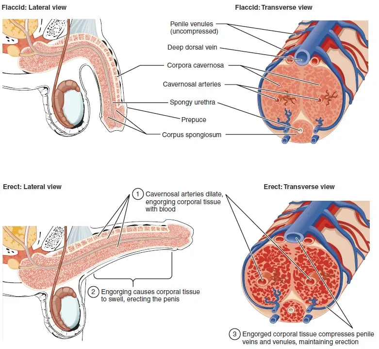
The male reproductive system is designed to produce, mature, and deliver sperm. The testes are the primary reproductive organs and are located within the scrotum. They contain numerous seminiferous tubules where sperm are produced through spermatogenesis.
The testes also produce testosterone, the primary male sex hormone. Testosterone regulates sperm production, drives development of secondary sex characteristics, and influences muscle mass, bone density, and libido.
Sperm Transport and Accessory Glands
Sperm produced in the testes move to the epididymis, a coiled tube where sperm mature and gain motility. During ejaculation, sperm travel from the epididymis through the vas deferens to the urethra.
Accessory glands contribute fluids that form semen. The seminal vesicles, prostate gland, and bulbourethral glands produce secretions that nourish sperm, protect them from acidic environments, and facilitate transport. These fluids increase semen volume and improve sperm survival during fertilization.
Penis and Erectile Function
The penis serves as the external organ for copulation and ejaculation and also acts as a conduit for urine. During sexual arousal, increased blood flow causes erection, allowing effective delivery of sperm into the female reproductive tract.
From an EMS perspective, understanding normal erectile physiology can help differentiate physiologic responses from pathologic conditions such as priapism or trauma-related vascular compromise.
Spermatogenesis: Producing Male Gametes
Spermatogenesis is the continuous process by which spermatogonia develop into mature sperm cells. This process begins at puberty and continues throughout life. It occurs within the seminiferous tubules of the testes and is regulated by follicle-stimulating hormone.
The process includes mitosis, meiosis, and spermiogenesis. Spermatogonia divide to form primary spermatocytes, which undergo meiosis to form haploid spermatids. Spermiogenesis then transforms spermatids into streamlined spermatozoa with tails for motility.
Millions of sperm are produced daily to ensure reproductive potential. Each sperm carries 23 chromosomes, contributing half of the genetic material required for fertilization.
Female Reproductive System: Structure and Function
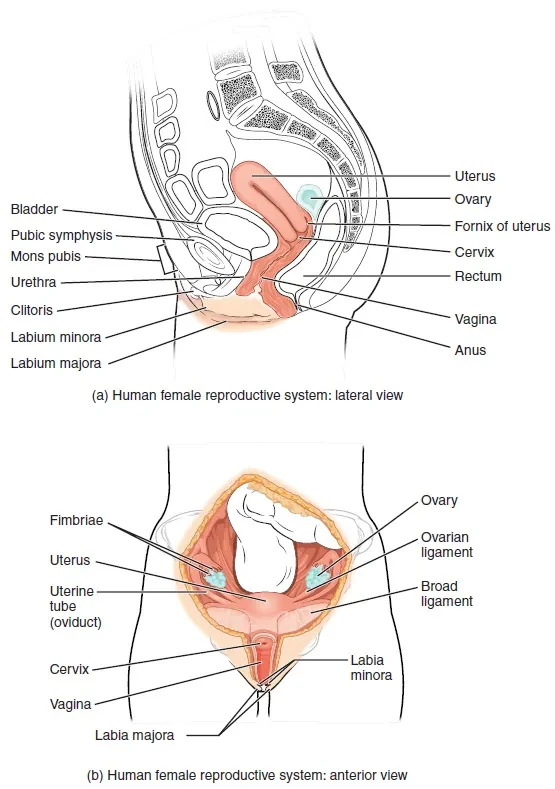
The female reproductive system produces eggs, supports fertilization, and provides an environment for fetal development. The ovaries are the primary reproductive organs and store ova. They also produce estrogen and progesterone, hormones essential for menstrual cycle regulation and reproductive health.
Each month, one mature egg is released from an ovary during ovulation. This cyclical process prepares the body for potential pregnancy.
Ovaries, Fallopian Tubes, and Fertilization
Following ovulation, the released egg is captured by finger-like structures called fimbriae and guided into the fallopian tubes. Fertilization typically occurs within the fallopian tubes if sperm are present.
Once fertilized, the resulting zygote travels toward the uterus aided by smooth muscle contractions and ciliary motion. This journey is critical for successful implantation and early embryonic development.
Uterus, Cervix, and Vagina
The uterus is a pear-shaped muscular organ where implantation occurs and fetal development is supported. It consists of the fundus, body, and cervix. The cervix forms the lower portion of the uterus and acts as a protective barrier against infection while allowing sperm passage.
The vagina is a muscular canal approximately 10 centimeters long. It serves as the entrance to the reproductive tract and functions as the birth canal during delivery.
Oogenesis: Producing Female Gametes
Oogenesis is the process by which female precursor cells develop into mature eggs. Unlike spermatogenesis, oogenesis begins during fetal development. Primary oocytes form and remain arrested in the first stage of meiosis until puberty.
At puberty, ovulation begins. Each month, luteinizing hormone triggers completion of the first meiotic division, producing a secondary oocyte and a polar body. The cytoplasm divides unequally, ensuring the surviving egg has sufficient resources for early development.
If fertilization occurs, the secondary oocyte completes meiosis and becomes a mature ovum. When fertilized by a haploid sperm, a diploid zygote with 46 chromosomes forms.
The Menstrual Cycle: Coordinated Ovarian and Uterine Cycles
The menstrual cycle consists of two interrelated cycles. The ovarian cycle describes changes within the ovaries. The uterine cycle describes changes in the uterine lining. Together, these cycles prepare the body for possible pregnancy and typically span about 28 days.
Ovarian Cycle Phases
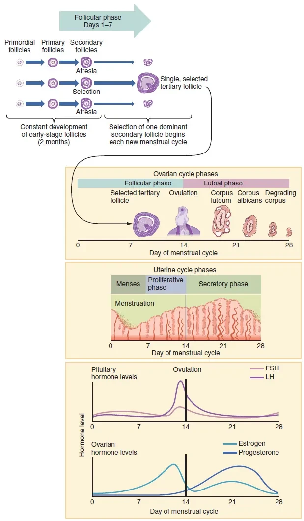
The ovarian cycle has two main phases. The follicular phase begins the cycle and is driven by follicle-stimulating hormone. Multiple follicles begin to mature, but typically only one becomes dominant. Rising estrogen levels from the dominant follicle stimulate uterine lining growth.
Ovulation occurs following a surge in luteinizing hormone. After ovulation, the luteal phase begins. The ruptured follicle becomes the corpus luteum, which secretes progesterone to support the uterine lining. If fertilization does not occur, the corpus luteum degenerates and hormone levels fall.
Uterine Cycle Phases
The uterine cycle includes the menses, proliferative, and secretory phases. Menses marks the shedding of the uterine lining due to declining progesterone levels. This phase results in menstrual bleeding.
The proliferative phase follows, during which estrogen stimulates regeneration and thickening of the endometrial lining. The secretory phase aligns with the luteal phase and prepares the uterus for implantation. If fertilization does not occur, the lining is shed and a new cycle begins.
Clinical Reinforcement for EMTs
Reproductive anatomy and physiology influence many EMS encounters. Menstrual history can provide clues to pregnancy status. Hormonal changes can affect vital signs, fluid retention, and mood. Understanding fertilization and implantation helps frame ectopic pregnancy risk and early pregnancy complications.
Clear, respectful communication about reproductive health is essential in patient assessment. Using correct terminology and understanding normal physiology improves accuracy, professionalism, and patient trust.
Critical Thinking Questions
A patient reports abdominal pain and missed menses. How does your understanding of ovulation and implantation influence your suspicion for early pregnancy or ectopic pregnancy?
How do hormonal changes during the menstrual cycle influence fluid balance and symptom reporting in some patients?
A patient presents with pelvic pain shortly after ovulation. How could knowledge of ovarian and uterine cycle timing help guide your assessment and communication with the receiving facility?
Why does oogenesis produce only one viable ovum per cycle while spermatogenesis produces millions of sperm daily, and how does this affect fertilization probability?
How does understanding the function of the cervix and uterus support patient education and reassurance during non-emergent reproductive complaints?
Image Credits: Image from Anatomy and Physiology by OpenStax, licensed under CC BY 4.0